Showing posts with label microbiology. Show all posts

Wednesday, October 30, 2013

microscopic crafts

A Saturday shopping adventure in Melbourne with my friend Mimi brought us to a cabinet of medical curiosities, tucked away in the magical Nicholas Building. Anno Domni Home is filled with sculptures which celebrate the beauty in the macabre. My favourite, some petri dishes of micro-organisms, made from hospital blankets.


There are also embroideries, felted body parts and other stitched objects which will fascinate anyone with a medical or scientific interest. Considering that Mimi and I bonded in medical school over dissection classes, sewing and imagining microbiology lecture slides as fabric patterns, it is no wonder that our wanderings brought us to this crafty cave. Watch out for an exhibition next year by this clever artist.

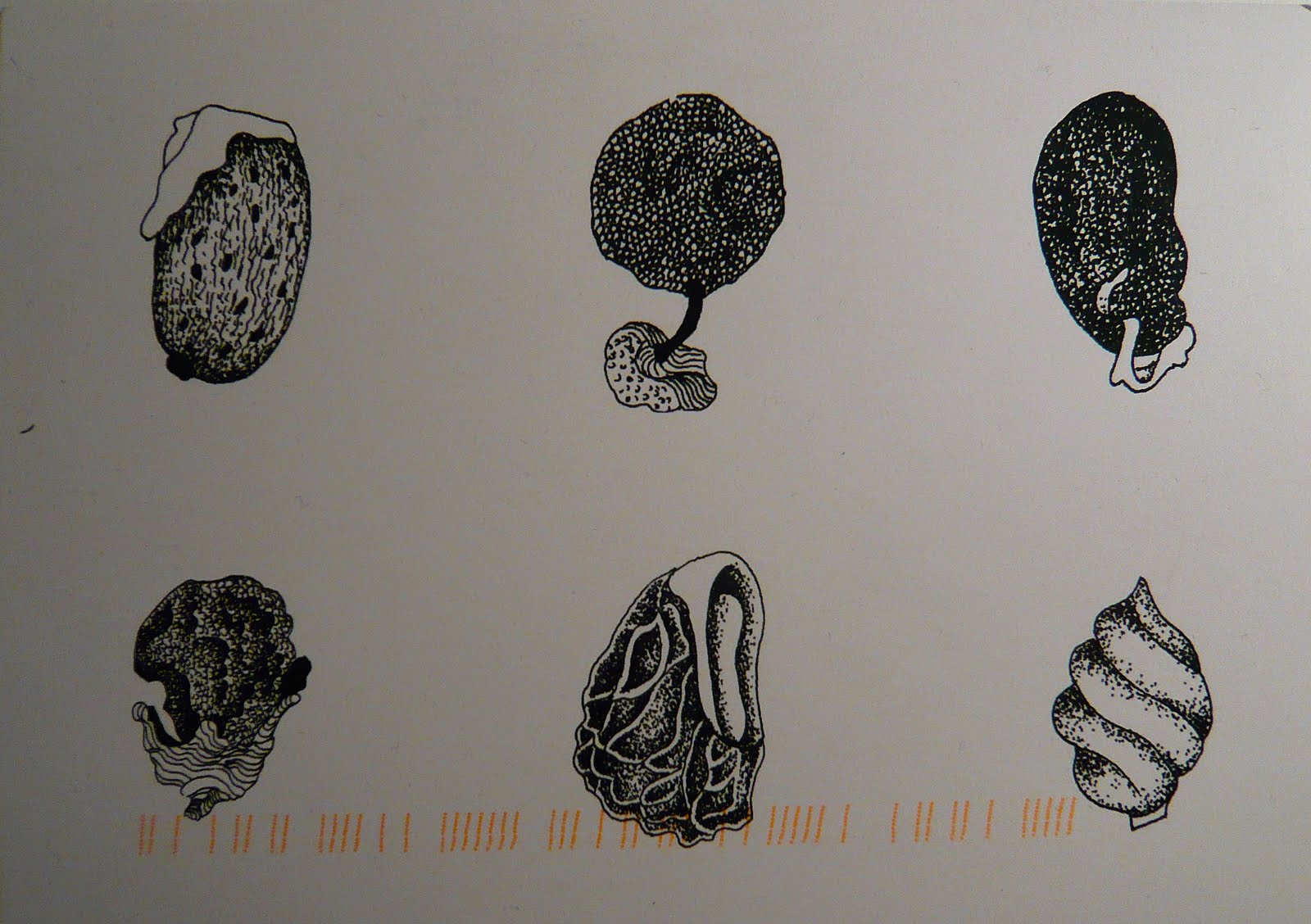
Image from Anno Domini Home blog, used with kind permission of Andrew Delaney.

Wednesday, March 21, 2012

as winter slides into spring

It seems only fitting to mark the changes of the seasons with some beautiful microscopic slides. Two years ago I was fascinated by the nonist's exquisite microscopic specimens. Now I have discovered Wilson Bentley's 1880s microscope-photographs of the ephemeral, melting, delicate snowflake:


Images from Wikimedia Commons.

Thursday, February 2, 2012

stitching seed cells

Following a previous post about Anna Dumitriu's experiments in bacterial communication, I thought I would share the embroidery I finished last year, inspired by Annie's postcard of seed cells. More about pneumatic tubes next week I promise!


Tuesday, August 2, 2011

experiments in bacterial communication

Another post not strictly about pneumatic transportation, but on a topic that is closely (or perhaps more tangentially) related if we think of the ways in which pathological samples are networked around a hospital as a form of communication.


This is a piece of embroidery by Anna Dumitriu. Anna is interested in the borderlines between art and science, such as this example of bacterial communication, tracing the movement of bacterial cells on pieces of linen and lace. She is the director of the Institute of Unnecessary Research and is an artist in residence on the Modernising Medical Microbiology Project at Oxford University. Her exhibition of these exquisite embroideries opened on Saturday at R-Space, in Lisburn, Northern Ireland. The exhibition also includes other microbiological crafts such as:
"A large-scale collaborative crochet based on the bacteria from the artist’s own bed, an indigo blue coloured patchwork stained with MRSA bacteria grown on chromogenic agar and patterned with clinical antibiotics and other tools in the research and treatment of this disease, [and] a Whitework embroidered lab coat patterned with images of bacteria and moulds found on it"
Despite my interest in science, medicine, art and craft, I have not always been a fan of bioart, finding many works disengaging. This exhibition caught my eye however, not only for its melding of embroidery and microbiology (see my fascination with this in a previous post here and here), but also because of the beauty of each piece, and the curiosity the artist invokes in the viewer.


If you miss the exhibition you can always settle for the catalogue, available from Blurb.


Thursday, July 15, 2010

nonist's beautiful specimens

On previous posts, here and here, I have included photographs of vintage microscopic slides, but none have been as exquisite as the range that the nonist carefully collated on his blog, as sourced from a number of fantastic sites listed at the end of his post. I know that microscopic slides are not strictly related to pneumatic tubes, but perhaps there is some link between microscopic samples, pathology, Victorian science and medicine, handcrafted details or ...?


Images via the nonist.

Tuesday, June 29, 2010

my medical museum

The My Medical Museum competition has recently closed. I enjoyed looking at the other entries, which included several videos, and there are many of these museums that I would love to visit one day. Here is my entry for the competition:

I’ve always loved medical libraries, probably because I have spent over a decade studying in them. There are constant sources of distraction in medical libraries: the latest scientific journals; microbiology textbooks with exquisite photographs; anatomical texts filled with woodcuts and engravings. The library in my medical school in Tasmania, Australia was a rather modest affair but when I moved to the University of Melbourne in Victoria, Australia I found something much larger. My new medical library had multiple levels, rare old books, new computers and the most wonderful distraction of all: a medical museum.

Gradually, over the years, the Medical History Museum at the University of Melbourne has become more than a distraction, and since March this year I have been volunteering there every Thursday afternoon. I share a computer and little office with Ann (another staff member), an operating table, anaesthetic equipment and hundreds of locked and labelled wooden boxes. It is dusty and cluttered and I love it!

The museum was established in the library in 1967, with a grant from the Wellcome Trust. A beautiful 19th century Savory and Moore pharmacy, shipped from Belgravia, London, is installed in the museum, complete with bottles and gold-labelled herb drawers. On display there are also microscopes, amputation sets and bleeding equipment, in walnut display cases. Currently there is a temporary exhibition about apothecaries -The Physick Gardener: Aspects of an Apothecary's World - curated by the museum’s new curator Susie Shears. Behind a hidden door in the pharmacy are the curator’s offices and storage areas, where chests and drawers may contain pathological slides or stapleguns, and shelves are filled with boxes, books and ephemera.

There are many treasured items in the museum’s collection including specie jars, pill rolling machines and medicine chests used by doctors during visits to rural areas in Australia. One of the oldest photographs (1864), and one of my favourites, depicts the first medical students carrying out work in the Anatomy Dissecting Room, under the supervision of Professor Halford, and the watchful gaze of the medical school porter.

Professor George Britton Halford (1824 – 1910) was a lecturer in London, before taking the first chair of anatomy, physiology and pathology at the University of Melbourne. He moved to the antipodes with anatomical and pathological specimens he had collected for a museum, and books to start a library. His first practical classes and lectures were held in the converted coach-house of his private residence, before moving to the newly completed medical school in 1864.

Professor Halford played an important role in the teaching and administration of the new medical school in Melbourne, and was a strong advocate for female students. He arrived in Melbourne with an established record as a researcher (one of his most important essays being The Action and Sounds of the Heart: A Physiological Essay (1860)) but his later controversial experiments with snake venom damaged this reputation.

The objects and documents I found associated with Halford provide a window not only into the life of a contentious researcher and teacher, but also into the collection of the Medical History Museum. Amongst Halford’s material objects and paper artefacts, there is: a Powell and Lealand compound monocular and binocular microscope stored in a walnut case with a handwritten inventory; a cabinet of microscope slides commercially and handmade between 1860 and 1889; a paper entitled ‘On a Remarkable Symmetrically Deformed Skeleton’ (1868); photographs of the professor and his family; and his business card.

Other pieces in the collection associated with Halford include a student’s set of lecture notes compiled during Professor Halford’s anatomy and physiology lectures throughout 1877. This leather bound exercise book, with John Springthorpe’s scribblings and coloured pencil illustrations, is the only surviving example of Professor Halford’s teaching.

All of these objects are material remnants of Professor Halford’s time at the University of Melbourne. They are microscopic slices of a time when medical students wore aprons and dissected on wooden tables and when slides were handmade. The objects are portals into the past that overlap with my curiosity of the future. They are just some of the thousands of wonderful stories to be found in my medical museum.

All photographs are my own except for the anatomy class which was included with the kind permission of Susie Shears, Medical History Museum, University of Melbourne.

Thursday, June 3, 2010

seed cells


This card arrived by post several weeks ago from my friend Annie. They are the seeds of small Australian plants and Annie likened them the seed cells of humans, microbes, that I had embroidered onto napkins recently. Her paper post gave me the idea to continue embroidering molecular representations of human life onto cloth, as part of an ongoing exploration of the relationship between new medical technologies and old, between the digital and the stitched.

Tuesday, June 1, 2010

pandemic!

On the weekend I played Pandemic with Thomas and friends Ros and Jarrod. The timing could not have been better as I finish a draft of my report today for a larger project looking at the 'biosociality' of influenza pandemics. The game was fantastic. The virus spread in little coloured blocks all over the board, between cities, as we worked together, Medic, Scientist, Operations Expert and Researcher, to find cures for the disease. As we didn't actually eradicate the virus, it would have been more appropriate if we were finding vaccines, but small detail aside, the game was tense and exciting. Apparently new updates for the game include little petri dishes to store the virus! The game is out of stock at the moment, but as soon as it is reprinted in May/June, I'm ordering one ...


Image of green algae that I embroidered onto a napkin.